
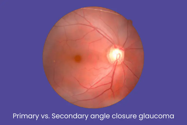
Primary vs. Secondary angle closure glaucoma
Glaucoma is one of the leading causes of irreversible blindness worldwide. Among its different types, angle closure glaucoma is especially serious, as it can cause sudden vision loss if not detected and treated early.
But not all angle closure glaucoma is the same. It is broadly classified into Primary and Secondary Angle Closure Glaucoma, and understanding the difference is key to proper diagnosis and treatment.
This blog aims to provide clear, patient-friendly information to help you understand how these two conditions differ, why they happen, and what treatments are available.
Table of Contents
ToggleWhat is angle closure glaucoma?
The eye produces a clear fluid called aqueous humor, which flows through the pupil and drains out via the drainage angle between the iris and the cornea. When this angle becomes blocked or narrowed, the fluid cannot drain properly, resulting in increased pressure inside the eye — a condition known as angle closure glaucoma.
If this pressure isn’t reduced quickly, it can permanently damage the optic nerve and lead to vision loss.
Primary Angle Closure Glaucoma (PACG)
")
What it is:
This form of glaucoma occurs without any other eye disease or external trigger. It usually develops due to anatomical characteristics of the eye, such as a shallow anterior chamber.
Symptoms:
- Sudden eye pain
- Blurred vision
- Halos around lights
- Headache
- Nausea and vomiting (especially during acute attacks).
Causes/Risk factors:
- Farsightedness (hyperopia)
- Naturally narrow angles.
- Aging (typically 40+)
- Female gender
- Family history of glaucoma.
- Asian ethnicity
Diagnosis:
- Gonioscopy (to examine the angle)
- Intraocular pressure (IOP) measurement.
- OCT and visual field tests.
Treatment:
Treatment goals:
- Relieve the block in fluid drainage.
- Prevent future attacks.
- Preserve optic nerve function and vision.
1. Laser Peripheral Iridotomy (LPI)
- What it is: A laser procedure that creates a small hole in the iris, allowing fluid to bypass the blocked angle and flow more freely.
- Why it matters: It’s usually the first-line treatment and often prevents recurrence of angle closure.
- Quick and painless, usually done as an outpatient procedure.
2. Medications (Before or After LPI)
- IOP-lowering drops:
- Beta-blockers (e.g., Timolol) – reduce fluid production.
- Prostaglandin analogs (e.g., Latanoprost) – increase fluid outflow.
- Carbonic anhydrase inhibitors (e.g., Dorzolamide) – decrease fluid production.
- Alpha agonists (e.g., Brimonidine) – reduce production & increase outflow.
- Beta-blockers (e.g., Timolol) – reduce fluid production.
- Oral medications (e.g., Acetazolamide) in acute cases to rapidly lower eye pressure.
3. Laser iridoplasty (if LPI is ineffective or angle remains narrow)
- Shrinks the peripheral iris using laser burns to widen the angle.
- Useful in eyes with plateau iris configuration.
4. Surgical Options (if necessary)
- Lens extraction: In some cases, early cataract surgery helps deepen the anterior chamber and open the angle.
- Trabeculectomy: A surgical drainage procedure if pressure is uncontrolled with medications or laser.
- Glaucoma drainage devices (implants): Used in complex cases.
Secondary angle closure glaucoma

What it is:
Unlike primary glaucoma, secondary angle closure glaucoma occurs due to another underlying eye condition or disease that interferes with the drainage angle.
Symptoms:
- Gradual or sudden vision changes.
- Eye pain
- Headache
- Symptoms of the underlying cause (e.g., redness, swelling).
Common causes:
- Inflammation (e.g., uveitis)
- Eye trauma or injury
- Lens-related problems (e.g., swollen cataract)
- Neovascularization (abnormal blood vessel growth)
- Tumors inside or near the eye.
Diagnosis:
- Detailed eye examination to detect underlying causes.
- Ultrasound or anterior segment imaging.
- Blood work if systemic diseases are suspected.
Treatment:
Treatment is more complex, as it involves managing both the high IOP and the underlying cause of the angle closure.
1. Treating the underlying cause
- Inflammation (e.g., Uveitis):
- Corticosteroid eye drops or oral steroids.
- Cycloplegic drops to relax the ciliary muscle.
- Swollen or displaced lens:
- Surgical removal of the lens (cataract extraction or lens repositioning).
- Surgical removal of the lens (cataract extraction or lens repositioning).
- Tumors or trauma:
- Targeted surgical or medical treatment depending on the diagnosis.
- Targeted surgical or medical treatment depending on the diagnosis.
- Neovascular glaucoma:
- Anti-VEGF injections to stop abnormal vessel growth.
- Pan-retinal photocoagulation (laser therapy) to reduce retinal ischemia.
2. IOP-lowering medications
- Similar to those used in primary angle closure.
- Often used alongside treatment for the root cause.
3. Laser or Surgical procedures
- Laser iridotomy or iridoplasty: If pupillary block is a contributing factor.
- Trabeculectomy or Glaucoma drainage implants: If angle cannot be opened or medication fails.
- Cyclodestructive procedures: In rare, end-stage cases to reduce fluid production by partially destroying the ciliary body.
Comparison table: Primary vs. Secondary angle closure glaucoma
Why is it important to act early?
Angle closure glaucoma can progress quickly and cause permanent vision loss if left untreated. Many people are unaware they have the condition until it’s too late. That’s why regular eye checkups, especially if you have risk factors, are essential.
If you notice any warning signs like eye pain, blurred vision, or halos around lights — don’t ignore them. Seek immediate medical attention.
📌 Important note: Never stop or adjust glaucoma medications without consulting your eye doctor. Sudden changes in eye pressure can damage vision permanently.
Conclusion
If you or your loved one is experiencing symptoms of glaucoma or has been diagnosed and needs guidance, expert care is just a call away.
Krisha Eye Hospital, Ahmedabad, offers comprehensive diagnostic and treatment services for all types of glaucoma, including laser and surgical options. Our experienced team uses advanced technology to preserve and protect your vision. Call us today to book an appointment or consultation.
Author bio
Dr. Dhwani Maheshwari, an esteemed ophthalmologist with over 10 years of experience, leads Krisha Eye hospital in Ahmedabad with a commitment to advanced, patient-centered eye care. Specializing in cataract and refractive surgery, Dr. Maheshwari has performed more than a thousand successful surgeries. Her expertise lies in phacoemulsification, a technique recognized for its precision in cataract treatment.
Dr. Maheshwari’s educational journey includes an MBBS from Smt. NHL MMC, a DOMS from M & J Institute of Ophthalmology, and a DNB in Ophthalmology from Mahatme Eye Bank Eye Hospital, Nagpur. She also completed a fellowship in phacoemulsification at Porecha Blindness Trust Hospital, further enhancing her surgical skills. In addition to her work at Krisha Eye Hospital, Dr. Maheshwari serves as a consultant ophthalmologist at Northstar Diagnostic Centre.
Under her leadership, Krisha Eye Hospital aims to bring all superspecialties under one roof, offering comprehensive eye care solutions for all vision needs.
FAQs
No, they are distinct conditions. However, complications or delayed treatment in primary glaucoma can lead to complex cases requiring more intensive management.
Both can cause permanent vision loss, but secondary glaucoma is often harder to manage because it’s linked to another underlying condition.
It is not curable, but it is manageable. Early diagnosis and treatment can help preserve vision and prevent progression.
If you’re over 40, have a family history, or are at risk, get a comprehensive eye exam at least once a year.
Untreated glaucoma can lead to sudden and irreversible blindness, especially in acute angle closure cases.
Yes, primary angle closure glaucoma often affects both eyes, though not always at the same time. Secondary glaucoma can be unilateral or bilateral depending on the underlying cause.
In acute angle closure, vision loss can occur within hours if left untreated. That’s why it’s considered an emergency and requires immediate medical attention.
Laser iridotomy is generally not painful. Some patients may feel a slight pinch or mild discomfort, but the procedure is quick and done under local anesthesia.
“Narrow angles” means there’s a risk of angle closure but it hasn’t happened yet. Regular monitoring or preventive treatment like prophylactic laser iridotomy may be recommended.
While no specific diet can prevent it, staying hydrated, avoiding certain medications (like decongestants), and regular eye check-ups can help reduce risk. However, anatomical predispositions can’t be changed through lifestyle.